
X-rays are electromagnetic radiation. It is a shorter wavelength than visible light. The rays are commonly used in medical imaging to visualize the inside of objects.
- Discovery of x-rays
- Properties of x-rays
- Production of X-rays
- What are X-rays used for? or types of x-rays
- What Happens During an X-Ray?
- what are the dangers of X-rays?
- Characteristics x -rays
- What is X-RAY TUBE?
- History of the developments of the X-ray tubes
- Principle of the X-ray tube
- Components of X-ray tube
Discovery of x-rays
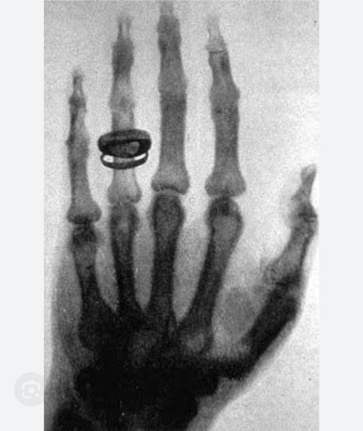
The first X-rays were discovered by Wilhelm Conrad Roentgen on Friday, November 8, 1895. Who was a German physicist, when they used Crooks tube. He called them ‘x-ray’s, ‘x’ for unknown. They were professor of physics at the University of Germany.
They were working on the cathode tube in his laboratory. Suddenly, he saw the shadow of the bone of his hand after the months he had published a carefully reasoned description of his work and the famous radiograph of his wife’s hand. It is an accidental discovery that marked the beginning of X-ray technology.
Properties of x-rays
- X-rays travel in a straight line.
- X-rays have electromagnetic waves.
- X-rays can’t travel in wavelength.
- X-rays have a wavelength from 0.01 to 10nm.
- X-rays can’t be focused by the lens.
- X-rays travel in a vacuum.
- X-rays have blackened in the photography.
- X-rays can’t be deflected by an electric field or magnetic field.
- X-rays are invisible rays.
- X -rays have energy from 100ev to 100kev.
Production of X-rays
X-rays are produced when fast-moving electrons are stopped using a target material. The moving electrons possess kinetic energy. When the electron is unexpectedly stopped, its kinetic energy is transformed into heat and x-rays. This conversation is taking place in the target material. Therefore, the interaction of electrons with the target is the basis for x production.
Electron interaction with the target
Ionization of target atoms
The fast-moving electron enters the surface layer of the target and undergoes collisions. This is the process of incident electron transfer of sufficient energy and removes an electron from the atoms. This involves small energy transfer, resulting in the ionization of target atoms. Interaction of electron with target atoms.(A) Ionization of target atoms.(B)

characteristics x -rays.(C) Interaction with nuclear field. (D) Interaction with the nucleus.
What are X-rays used for? or types of x-rays
X-rays are a form of electromagnetic radiation with wavelengths shorter than visible light. In the medical field, X-rays are commonly used to diagnose things like
- Broken bones
- Dislocated joints
- Arthritis
- Causes of abdominal pain, in some instances
- Cancer
- Tooth decay
Here are some other key applications of X-rays in healthcare:
Radiography (X-ray Imaging)
This is the most common use of X-rays. Radiography involves creating images of the inside of the body, typically to visualize bones and detect abnormalities. X-ray images are used for:
- Identifying fractures and dislocations.
- Examining the chest for conditions like pneumonia, lung cancer, and tuberculosis.
- Dental X-rays for assessing teeth and jaw structures.
Fluoroscopy
This real-time X-ray imaging technique is used for dynamic studies of internal structures. Fluoroscopy is employed in procedures such as:
- Barium swallow studies to examine the digestive tract.
- Angiography to visualize blood vessels.
- Orthopedic procedures for guided joint injections or placements of medical devices.
Computed Tomography (CT or CAT Scan)
CT scans use X-rays from different angles to create detailed cross-sectional images of the body. CT scans are useful for:
- Detecting and locating tumors.
- Imaging the brain for stroke or trauma assessment.
- Visualizing the chest and abdomen for various conditions.
Mammography
X-ray imaging of the breast is known as mammography. It is a crucial tool for:
- Breast cancer screening and early detection.
- Evaluating breast abnormalities and guiding biopsies.
LDL Cholesterol Symptoms, Cause, Treatment
Interventional Radiology
X-rays are used in minimally invasive procedures to guide medical interventions. Examples include:
- Angioplasty and stent arrangement for vascular conditions.
- Embolization to treat irregular blood vessels or block blood supply to tumors.
- Drainage procedures for fluid collections.
Bone Densitometry
X-rays are used to measure bone mineral density, assisting to diagnose conditions such as osteoporosis and assess fracture risk.
X-ray Crystallography
In research, X-rays are used to specify the atomic and molecular structure of a crystal. This is widely used in fields such as chemistry and biochemistry for analyzing the structure of proteins, nucleic acids, and other biological molecules.
Security Imaging
X-rays are utilized in security systems, such as airport baggage scanners, to notice objects that may pose a security hazard.
What Happens During an X-Ray?
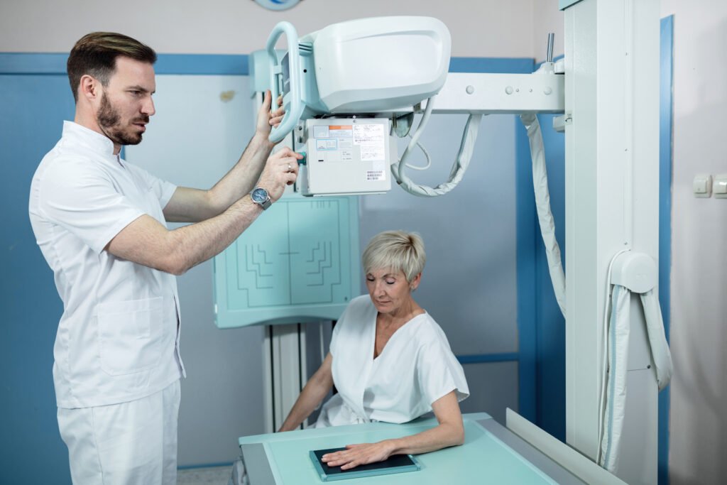
Most X-rays don’t require any special preparation. The doctor may ask you to take off jewelry, eyeglasses, a watch, or any metal objects or clothing that could get in the way of the image.
An X-ray expert can take images while you stand up or lie down. It depends on the part of your body being scanned. The X-ray tube hangs over the table. The film of X-rays is in a drawer under the table.
The machine transmits a beam of radiation via your body. Your rigid, dense bones restrict from entering that beam, so they show up as white on the black. The radiation also runs via softer tissue like fat & muscle, which occur in shades of gray in the X-ray. The air in your lungs will look black in the image.
The patient won’t feel anything during an X-ray, but it can be hard to hold still, and the examination table can be discomfiting for the patient. The x-ray technician may take images from a few various angles. They might use cushions or sandbags to prop up a body part to get a better view of the area. They’ll probably ask you to hold your breath till the image is captured so the image doesn’t blur.
Occasionally, the doctor needs more contrast in the image to see what’s going on for clear and authentic analysis. They might give you a contrast agent, like barium or iodine. You’ll either swallow it or bring it as a shot.
The machine makes clicks and buzzing sounds during the X-ray. The procedure could take just a few minutes for a bone X-ray or more than an hour for more complex problems.
what are the dangers of X-rays?
X-rays, like any form of ionizing radiation, carry potential risks, particularly when there is repeated or high-dose exposure. Here are some considerations regarding the dangers of X-rays:
Ionizing Radiation
X-rays are a form of ionizing radiation, which means they have enough energy to remove tightly bound electrons from atoms, potentially causing damage to biological tissues. Prolonged or excessive exposure to ionizing radiation can increase the risk of cellular damage and mutations.
Risk of Cancer
The primary concern associated with ionizing radiation susceptibility, including X-rays, is an increased risk of cancer. This risk is commonly considered to be accumulative over a person’s lifetime. Nevertheless, the risk depends on factors such as the dose of radiation, the age at exposure, and personal exposure.
Mental Health: Disorder, Symptoms, Diagnosis
Genetic Effects
High doses of ionizing radiation may generate modifications in the DNA of germ cells (sperm and eggs), potentially directing to hereditary genetic effects in offspring. This is a problem primarily at very high radiation doses, such as those encountered in particular occupational settings
Tissue Damage
In particular cases, high doses of radiation can cause direct damage to tissues, leading to radiation burns, radiation sickness, or other vital effects. However medical imaging techniques typically use low to moderate doses, underestimating the risk of immediate tissue damage
Pregnancy Concerns
Pregnant women are normally advised to minimize openness to ionizing radiation, particularly during the first trimester when the developing fetus is more sensitive to the effects of radiation. However, the risk-benefit balance is carefully assumed, and in some cases, the benefits of necessary diagnostic X-rays may outweigh the potential risks.
Multiple Sclerosis Cause, Symptoms, Diagnosis and Treatment
It’s important to highlight that the benefits of medical X-rays in diagnosing and monitoring health conditions often outweigh the possible risks. The use of X-rays in medical settings is carefully regulated, and healthcare professionals follow procedures to minimize radiation exposure to patients. Modern imaging equipment is developed to deliver the lowest effective dose of radiation required for accurate diagnosis.
Patients should speak openly with their healthcare providers about any worries they may have regarding X-ray procedures. Healthcare experts will consider the essentials of the imaging study and take suitable steps to ensure patient safety and minimize radiation exposure
Characteristics x -rays
Interaction of electron with target atoms
This is an interaction between the happening electron and the electron in the k shell. In this process, the happening electron hits the k shell, moves sufficient energy, and removes the k shell electron. The vacancy in the k shell is filled by an electron moving in words from the outer shell.
Interaction with the nuclear field
The incident electron occasionally reaches nearer to the nucleus of an atom in the target. Since the electron is a negative particle, it is attracted by the positive nucleus. It is made to orbit partially around the nucleus, decelerates, and goes out with reduced energy.
Interaction with nucleus
The electron may hit the nucleus directly and is stopped completely in a single collision.
In general, the interaction of B, C, and D are very rare in the diagnostic range of energies, leading to a lesser amount of X-ray production. The ionization collision dominates (99%) the interaction process and produces atoms.
What is X-RAY TUBE?
Introduction
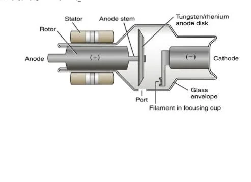
- An X-ray tube is a device that is meant for the production of X-rays.
- An X-ray tube is a vacuum tube.
- Contains a pair of electrons i.e.Cathode, an Anode
Cathode
The cathode is a filament that releases electrons when high voltage is applied. The cathode is a negative charge.
Anode
The anode is made up of tungsten, and the anode charge is positive. The anode attracts the electron.
- The electrons released from the Cathode come in contact with the tungsten, they release energy in the form of photons.
- These highly energized photons are channeled through a lead cylinder and a series of filters creating X-rays.
History of the developments of the X-ray tubes
- In 1895: x-rays were discovered from an experimental discharge tune called as crooks tune and cold cathode tube.
- In 1913: Crooks tube was improved by W.C c/as Coolidge tube or hot cathode tube.
- In 1915: Hooded anode tube.
- In 1920: an oil-cooled tube was discovered.
- In 1929: Rotating anode tube.
- In 1932: Grid-controlled stationary anode tube.
- In 1937: Grid-controlled rotating anode tube.
- In 1959: High school speed tune.
- In 1962:Rhenium alloyed tungsten composite anode tube.
- In 1967: Mammography unit with Mb anode.
- Principle of the X-ray tube
Principle of the X-ray tube
- An X-ray tube is a device based on the principle of energy conversion.
- In this process kinetic energy of a fast-moving electron is lost and converted into heat (99%) and x energy (1%).
- Made up of a small plate of tungsten 2 or 3 mm thick that is embedded in a large mass of cu.
- We use Tungsten as a target material.
- High atomic no.(74)
- High melting point (3370°c )
- It doesn’t vaporize easily.
Components of X-ray tube
- Cathode.
- Filament.
- Tube housing.
- Focusing cup.
- Glass Envelope.
- Focal spot.
- Anode.
- Stationary Anode.
- Rotating anode.
Cathode
The cathode is a negative charge in an X-ray tube. It consists of a metal structure to support the filament. It is the source of electrons.
Filament
It is a coil wire-like structure. It is 2mm in diameter and 1-2 cm long. It is made up of tungsten. Filament emits electrons when it is heated.
Tube housing
It is a protective casing that surrounds the x-ray tube to shield against radiation exposure and enhance safety. Protect against electric shock.
Focusing cup
It is embedded in a metal cup known as a focusing cup. The filament is a negative charge. It is made up of molybdenum and nickel.
Glass Envelope
x-ray tube is a vacuum tube. It contains the cathode and anode. It is 30 to 50 cm long and 20 cm in diameter. The glass Envelope is made up of Pyrex glass.
Collimator
It is a device that shapes and directs the x-ray beam to the area of interest, reducing unnecessary radiation exposure to adjacent tissues.
Focal spot
The area on the anode where the electron beam strikes, determines the size and sharpness of the x-ray image.
Anode
The anode is the positive electrode. It serves as the target for the electron beam converting kinetic energy into x-rays. It is made up of tungsten and copper. There are two types of anode
Stationary anode
- It is used in dental & portable x-ray machines.
- It is made up of tungsten and copper anode.
- It has low electric power.
- The production of X-rays at low or medium intensities.
- It conducts heat away from the focal spot.

Rotating anode
In 1993, the rotating anode tube was invented.
- It is used in general x-ray tubes.
- It is made up of tungsten+ Rhenium (5 %) as it provides mechanical strength.
- In this tube, the anode is made to rotate before the electron i emitted.
- It was developed to increase the heat loading with higher x-ray output.
- In these tubes, the electrons transfer their energy over a large area of a rotating target.
- Rotating anode tubes are large.